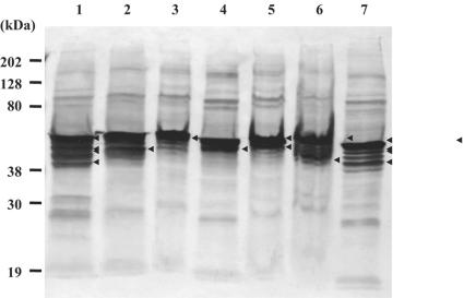
FIG. 4.
Expression profiles of vimentin in different cell lines by Western blot analysis. The total cellular proteins were extracted from each cell line and separated by SDS-PAGE (lane 1, MARC-145; lane 2, BHK-21; lane 3, CRFK; lane 4, MDCK; lane 5, PK-15; lane 6, ST; lane 7, Vero). The proteins were transferred onto a nitrocellulose membrane and incubated with monoclonal anti-vimentin Ab, followed by incubation with peroxidase-labeled anti-mouse IgG (H+L). The protein bands were detected by the addition of TMB membrane peroxidase substrate (one component). The major bands are indicated by arrowheads.

