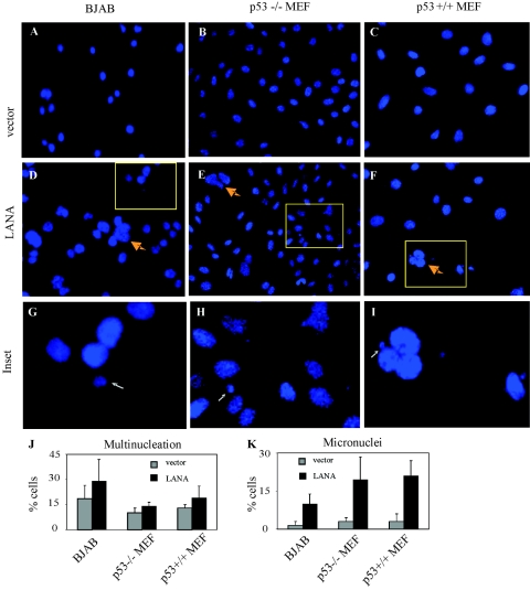
FIG. 4.
Induction of multinucleation and micronuclei in LANA-expressing cells. BJAB cells, p53−/− MEF, and p53+/+ MEF were transfected with a LANA construct and subjected to G418 selection. (A-C) DAPI staining of BJAB cells (A), p53−/− MEF (B), and p53+/+ MEF (C) with mock control DNA. (D-F) DAPI staining of representative fields shows increased multinucleation (yellow arrows) and numbers of micronuclei (white arrows) in BJAB cells (D), p53−/− MEF (E), and p53+/+ MEF (F) with LANA stably expressed. (G-I) Inset fields show micronuclei (white arrows) corresponding to the yellow rectangles in panels D to F. (J-K) Quantification of cells with more than two nuclei (J) and micronuclei (K).

