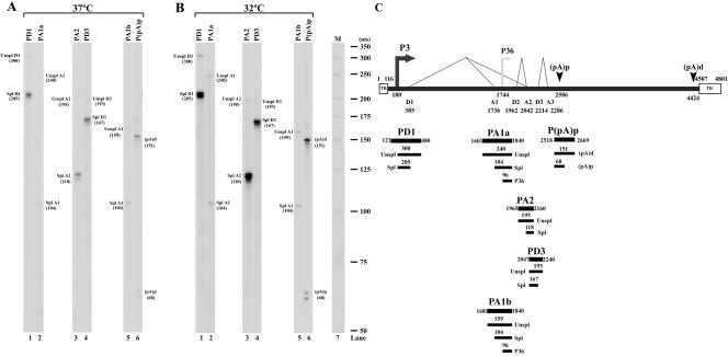
FIG. 1.
RNase protections of AMDV RNA. CRFK cells were infected with AMDV-G at 37°C (A) and 32°C (B). Total RNA isolated 6 days after infection was used for RNase protection assay. The probes are indicated at the top of each lane, and the identity of the RNA species protected by each band is indicated to either the right or the left. The probes are diagrammed in panel C, in relation to the previously published map of AMDV, with the predicted protected bands shown below each probe. The promoters are shown by arrows and the splice junctions by thin lines. Donor and acceptor sites are indicated, and the P36 promoter is depicted as a gray arrow to indicate that its presence is not confirmed by the experiments in the manuscript. A 32P-labeled RNA ladder (32) with the respective sizes indicated to the left is presented in panel B, lane 7. Unspl, unspliced; Spl, spliced; TR, inverted hairpin terminus.

