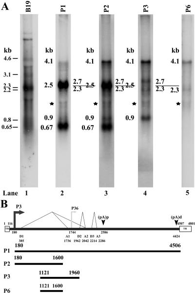
FIG. 2.
Northern analysis of RNA generated during AMDV-G infection of CRFK cells at 37°C. Total RNA isolated from AMDV-G-infected CRFK cells 6 days postinfection at 37°C was used for Northern analysis. RNA from uninfected CRFK cells was also assayed as a control and showed no hybridization (data not shown). (A) The sizes of the bands are shown to the left of each panel and are described in the text and in the legend for Fig. 3. RNA from B19-transfected COS cells, hybridized with a B19 genomic clone, was used for marker purposes (31) (lane 1). The probes used for hybridization are shown at the top of each lane. The bands indicated by the stars in lanes 2 to 5 run at the borders of the abundant unlabeled ribosomal RNAs and are likely to represent radioactivity excluded from this area of the gel. (B) The probes are diagrammed with respective nucleotide numbers and are shown in relation to the previously published map of AMDV, as described in the legend for Fig. 1. TR, inverted hairpin terminus.

