FIG. 4.
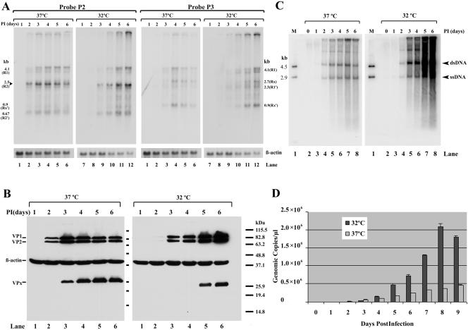
Time course of viral macromolecular synthesis following AMDV-G infection of CRFK cells at 32°C and 37°C. CRFK cells were infected with AMDV-G at 37°C or 32°C as indicated, and total RNA, DNA, capsid proteins, and generated virus were isolated at daily intervals. (A) Northern blot with probe P2 or P3 as indicated. The identities of the transcripts are shown to the left or right of the panels. R2 increased coordinately with capsid protein production (probe P2), while no RNAs initiated at P36 were detected (probe P3). Accumulation of β-actin RNA is shown as an internal control. (B) Western blot analysis showing accumulation of capsid proteins VP1 and VP2 at the time points and temperatures indicated. The accumulation of β-actin is shown as an internal control. VPx is an uncharacterized band that is likely a breakdown product of the capsid proteins. Markers are shown to the right. (C) Accumulation of replicating AMDV DNA at the times and temperatures shown. The replicative intermediates (double-stranded DNA [dsDNA] and single-stranded DNA [ssDNA]) are indicated. Lane 1 contains DNA markers of 4.5 and 2.9 kb. (D) Accumulation of AMDV virus as determined by quantitative PCR at either 37°C or 32°C at the indicated times postinfection.

