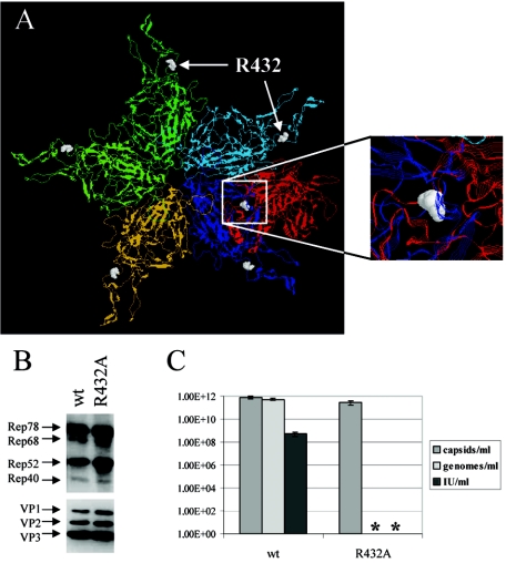
FIG. 3.
Characterization of the R432A packaging mutant. (A) A backbone model of a VP pentamer and a neighboring dimer is shown from the outside of the capsid; the subunits are shown in different colors (the pentamer is shown in cyan, blue, orange, and green; the dimer is shown in cyan and red) and amino acid R432 is represented as a space-filling model shown in white. The inset shows the R432 of the red subunit of the dimer interacting with the blue subunit of the pentamer. Viral supernatants were generated from 293T cells transfected with an AAV2 wt or mutated genomic plasmid. Freeze-thaw supernatants were assayed for viral protein expression (Western blot analysis using monoclonal anti-Rep [303.9] or anti-VP [B1] antibodies) (B) or ELISA-based AAV2 capsid titer, quantity of encapsidated AAV2 DNA, and infectious viral titer (C). Means ± standard deviations from at least four independent experiments are shown; an asterisk indicates that neither genomes nor infectious particles were detected for the R432A mutant. IU, infectious units.

