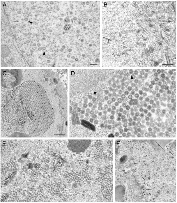
FIG. 8.
Ultrastructural analysis of insect cells infected with viruses expressing VP19C TN mutants. Sf9 cells were infected and the cells were processed for conventional EM as described in the legend of Fig. 4. Assembled capsids were evident in cells infected with the viruses expressing the wild-type proteins, indicated by black arrowheads (A), as well as for those with transpositions at amino acids 179 (C and D) and 443 (E). Capsids were not detected in any of the sections examined for cells infected with viruses expressing transposition insertions at amino acids 331 (B) and 237 (F). Bars, 0.2 μm (A and D), 0.5 μm (B, E, and F), and 1.0 μm (C).

