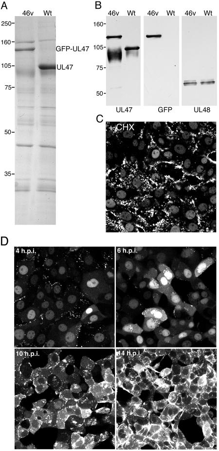
FIG.2.
Characterization of BHV-1 expressing GFP-UL47. (A) Equivalent amounts of extracellular virions purified from Wt- and jv46v-infected MDBK cells were analyzed by SDS-PAGE followed by Coomassie blue staining. (B) The same virions were analyzed by Western blotting with antibodies against UL47, GFP, and UL48. (C) MDBK cells grown in a coverslip chamber were infected with jv46v at a multiplicity of 10 in the presence of 100 μg/ml cycloheximide. One hour later, the cells were imaged on a Zeiss LSM410 confocal microscope. (D) MDBK cells grown in a coverslip chamber were infected with jv46v at a multiplicity of 2. At various times after infection, representative images were collected using a Zeiss LSM410 confocal microscope.

