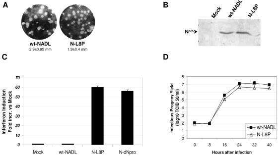
FIG. 6.
IFN-α/β induction by L8P Npro mutant BVDV. (A) Plaque morphology of N-L8P mutant compared to wt NADL. Bovine testicle cells were infected with the different viruses for 4 days at 37°C. Plaque formation was revealed by crystal violet staining. Mean plaque diameters ± standard errors of the mean are shown. (B) Western blot analysis of Npro protein expression and processing. Lysates from cells infected for 20 h were analyzed in Western blots probed with anti-Npro polyclonal antibody. The migration of the Npro protein is indicated. (C) IFN production by cells infected with Npro mutant BVDVs. Cell culture media from cells infected with N-L8P at an MOI of 10 for 24 h were harvested and assayed for IFN in the ISRE reporter cell line as indicated above. The induction was calculated relative to the values from mock-infected cells. The error bars indicate standard deviations. (D) Growth curve of wt NADL and N-L8P viruses. Bovine uterine cells seeded in 35-mm dishes (5 × 10⁵ cells/dish) were infected at a multiplicity of infection of 0.3. Culture media were harvested at appropriate times and cleared by centrifugation, and the infectivity was determined by endpoint dilution as described in Materials and Methods.

