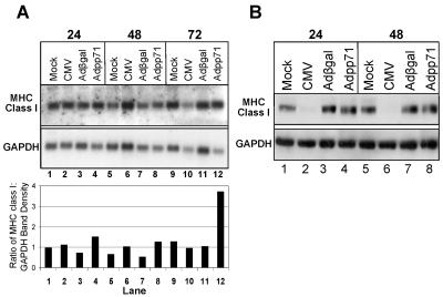
FIG. 5.
MHC class I transcript and protein levels are not altered in cells expressing pp71. (A) Autoradiographic and film images of electrophoretically separated RNA hybridized to HLA-B7-specific and GAPDH-specific probes. Replicate cultures of U373:CIITA cells in six-well plates were mock infected, exposed to 15 PFU/cell of the AD169 strain of CMV, or exposed to 100 PFU/cell of recombinant adenoviruses containing either the β-galactosidase gene or UL82. Cells exposed to virus were harvested at the times indicated. The cells of one well for each infection were solubilized, total RNA was isolated, and equivalent amounts of RNA were subjected to electrophoresis in formaldehyde-agarose gels in duplicate. RNA was transferred to nylon membranes by positive pressure. One nylon blot was incubated with 32P-labeled cDNA for HLA-B7 and the duplicate blot with a digoxigenin-labeled cDNA probe for GAPDH as described in Materials and Methods. 32P-labeled probes hybridized to MHC class I transcripts were visualized by autoradiography. Digoxigenin-labeled probes hybridized to GAPDH transcripts were reacted with anti-DIG antibody and visualized by chemiluminescence. Integrated optical density determinations were made for each relevant transcript, and the ratio of MHC class I to GAPDH transcript density is indicated for each lane below the autoradiographic and film images. (B) Film image of electrophoretically separated cell lysates reacted with antibody to MHC class I and GAPDH. To assess MHC class I protein levels, cells from the second well for each infection were harvested and solubilized and equivalent amounts of proteins were subjected to electrophoresis in a denaturing acrylamide gel. Proteins were transferred to nitrocellulose sheets and reacted with antibodies to MHC class I and GAPDH as described in Materials and Methods.

