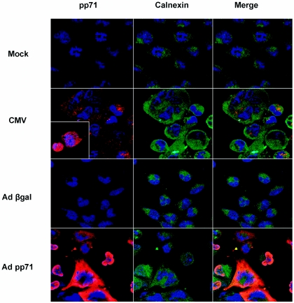
FIG. 7.
Nuclear and cytosolic localization of pp71. Immunofluorescence images of pp71 and calnexin in infected cells. Cultures of U373:CIITA cells cultured on glass coverslips in six-well plates were mock infected or exposed to 50 PFU of the AD169 strain of CMV per cell or to 100 PFU of Adβgal or Adpp71 recombinant adenoviruses per cell. At 48 h after infection cells were fixed and stained with antibody to pp71 followed by rhodamine-conjugated secondary antibody, and subsequently stained with FITC-conjugated anticalnexin antibody. Nuclei were visualized with DRAQ 5. Images of pp71 reactivity are shown in the left column, images of calnexin reactivity are in the middle column, and merged images are shown in the right column. The inset image of CMV-infected cells shows pp71 reactivity in nuclear and cytosolic compartments at this time point.

