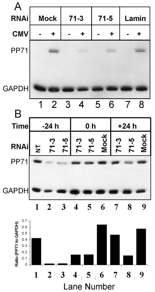
FIG. 9.
RNAi-mediated knockdown of pp71 protein accumulation. Film image of electrophoretically separated cell lysates reacted with antibody to pp71 or GAPDH. (A) U373:CIITA cells cultured in 24-well plates were transfected with 0.84 μg of the indicated RNAi duplex per well. Twenty-four hours after transfection, cells were mock infected or exposed to 15 PFU of AD169 per cell. Cells were harvested 48 h after infection and solubilized, and equivalent amounts of proteins were subjected to electrophoresis in a denaturing acrylamide gel. Proteins were transferred to nitrocellulose sheets and reacted with antibodies to MHC class I and GAPDH as described in Materials and Methods. (B) Cells were left untreated, exposed to transfection reagent only (mock), or transfected with 0.84 μg of 71-3 or 71-5 RNAi duplex 24 h before infection (−24 h), immediately after removal of the virus inoculum (0 h), or 24 h after infection (+24 h). Protein lysates were generated and analyzed as described for panel A. Integrated optical density determinations were made for pp71 and GAPDH signals, and the ratio of pp71 to GAPDH protein density is indicated for each lane below the film image.

