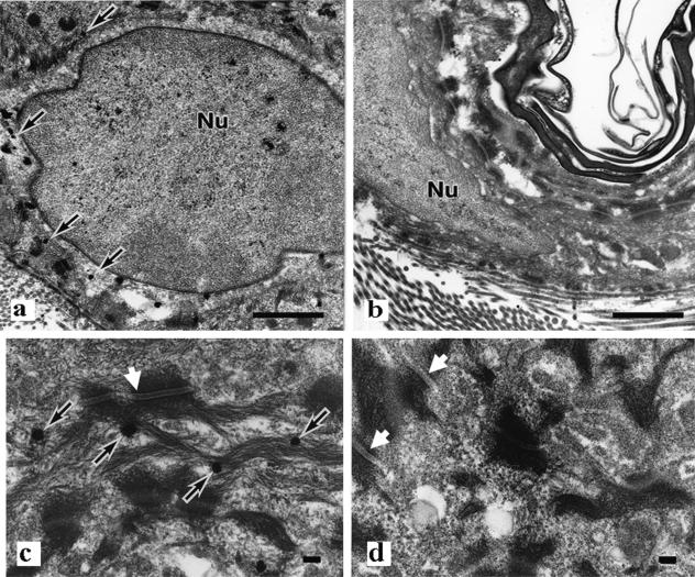
FIG. 3.
Electron micrographs of keratinocytes from the dorsal epidermis with low (a and b) and high (c and d) magnification. There are no apparent abnormalities in the keratin filaments and desmosomes (white arrows) of wild-type mice (a and c) and EPPK−/− mice (b and d). Silver grains are visible adjacent to keratin bundles (black arrows). Nu, nucleus. Bars = 1 μm (a and b) and 100 nm (c and d).

