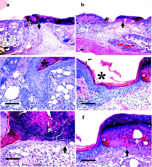
FIG. 5.
Histological analysis of skin wounds on the backs of wild-type (a, c, and e) and EPPK−/− (b, d, and f) mice. On day 4, the edges of wounds in wild-type mice invaginated sharply, and the epidermis was hypertrophic at the margins (a). A large scab covered the ulcer. In EPPK−/− mice, there was decreased hypertrophy of the epidermis at the wound's edge, and the leading edge extended a long distance compared with that in wild-type mice. The scab was also smaller than that in wild-type mice (b). *, wound edge; arrows, leading edge. Higher-magnification views of the wound edge (c and d) and of the leading edge (e and f) are shown. Bars = 100 μm.

