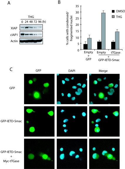
FIG. 7.
XIAP and cIAP-1 are decreased during THG treatment, and tTGase compensates for the lost IAP function for cell survival. (A) HCT116 Bax−/− cells were exposed to 1 μM THG for the indicated periods of time and subjected to immunoblot analysis with antibodies specific for β-actin, XIAP, or cIAP-1. (B and C) HCT116 Bax−/− cells were transfected with GFP, GFP-IETD-Smac, or GFP-IETD-Smac plus Myc-tTGase expression plasmids and treated with 1 μM THG for 48 h. The cells were stained with DAPI and analyzed under a fluorescence microscope (C), and GFP-positive cells with condensed or fragmented nuclei were counted as apoptotic cells (B). DMSO, dimethyl sulfoxide.

