FIG. 4.
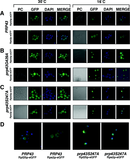
Both small- and large-ribosomal-subunit proteins exhibit nucleolar and nuclear accumulation in prp43 mutant cells at nonpermissive temperatures. (A) Phase-contrast (PC), GFP, DNA strain (DAPI [4′,6′-diamidino-2-phenylindole]), and merged images of GFP plus DAPI (MERGE) from PRP43 cells grown at the permissive temperature (30°C; left panel) or nonpermissive temperature (16°C; right panel) and containing the Rpl25p-eGFP reporter construct (top row) or the Rps2p-eGFP reporter construct (bottom row). Images similar to those described above (A) using a strain containing the prp43G429A mutation are shown in panel B, and those using a strain containing the prp43S247A mutation are shown in panel C. (D) The merged magnified images of the Rpl25p-eGFP and Rps2p-eGFP reporter strains are shown from the nonpermissive temperature conditions for PRP43 (left panels) and prp43S247A (right panels) to highlight the nucleolar and nuclear accumulation of the ribosomal subunit proteins.

