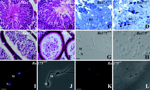
FIG. 4.
Spermatogenesis at stage VIII and germ cells in the cauda epididymidis of Ra175+/+ and Ra175−/−. (A, C, E, G, I, and J) Ra175+/+; (B, D, F, H, K, and L) Ra175−/−; (A, B, E, and F) hematoxylin and eosin (HE) staining; (C and D) toluidine blue staining; (G and H) Nomarski images; (I and K) fluorescence images with Mitotracker staining for mitochondria (red) and Hoechst staining for the nucleus (blue); (J and L) bright-field images. Compared to normal spermatogenesis in Ra175+/+ testes (A and C), spermatogenesis normally proceeds until the round spermatid stage (B and D), but abnormally shaped germ cells appear in elongating stages (B) in Ra175−/− testes. In contrast with Ra175+/+ testes (A and C), the areas for the residual bodies and the thick portions of spermatid tails (mitochondrial regions indicated by asterisks) are almost absent in Ra175−/− testes (B and D). In contrast with Ra175+/+ (E, G, I, and J), mature spermatozoa are not detected, but many exfoliated germ cells are detected in the cauda epididymal lumen of Ra175−/− (F, H, K, and L). Note that thick portions (mitochondrial regions) are absent in the exfoliated elongated spermatid tails in the Ra175−/− (H, K, and L). Thus, immature elongated spermatids in the Ra175−/− cauda epididymidis lacked mitochondrial region, which eventually leads to sterility. RS, round spermatids; M, middle piece; N, nucleus. Arrows indicate irregular nuclei. Bars in panels A, B, E, and F, 20 μm. Bars in panels C and D, 5 μm. Bars in panels G to L, 10 μm.

