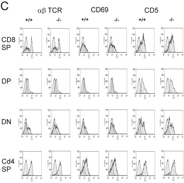
FIG. 2.
Normal differentiation in thymuses of wild-type and Rap1A−/− mice. (A) Expression pattern for CD4 and CD8. (B) CD44 and CD25 expression in CD4− CD8−population. Thymocytes were four-color stained for CD4, CD8, CD44, and CD25 markers. The results are representative for one of 10 sets of wild-type and Rap1A mutant mice. (C) Positive selection in the thymus is intact in Rap1A−/− mice. The thymus was three-color stained for CD4 PE, CD8 APC, and FITC-labeled αβTCR, CD69, or CD5. Expression profiles for each FITC marker were measured in each CD4/CD8 population.


