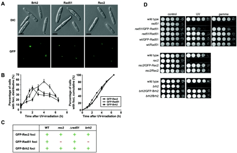
FIG. 1.
Focus formation. Wild-type (WT) (UCM350) cells expressing GFP-Rad51, Rec2, or Brh2 were viewed by differential interference contrast (DIC) imaging and fluorescence microscopy without fixation. (A) Cells were examined for focus formation 4 h after irradiation with UV (30 J/m2). Cells with representative Brh2, Rad51, or Rec2 foci are shown. Typically, cells had one to two foci. Bar indicates 3 μm. (B) The fraction of cells with foci at each time point was determined after counting approximately 200 cells. The cumulative representations of each distribution are shown on the right. (C) rec2 (UCM54), brh2 (UCM565), or rad51 (UCM628) mutant cells expressing GFP-Rad51, Rec2, or Brh2 were examined for foci at 2 and 4 h as described above. +, competent in focus formation; −, no focus formation. (D) Survival of rad51, rec2, and brh2 mutant strains expressing GFP-tagged or untagged Rad51, Rec2, or Brh2, respectively, after irradiation with UV (120 J/m2) and, in the case of rad51, with gamma rays (400 Gy). Serial 10-fold dilutions of cell suspensions were spotted from left to right as shown.

