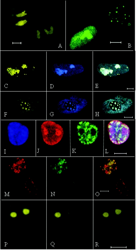
FIG. 2.
Confocal images of nuclei of transfected cells. Bars, 5 μm. (A) Meq-EYFP distributed throughout nuclei and nucleoplasm of two cells. (B) Meq/vIL8-EYFP associated with Cajal bodies (punctate) and nucleus in cell at upper right and with the nucleoplasm and nucleolus in the cell at bottom left. (C to E) Cell cotransfected with Meq-ECFP and Meq/vIL8-EYFP. (C) Meq/vIL8-ECFP exhibiting nucleoplasmic, nucleolar, and Cajal body association. (D) Meq-ECFP showing association with the nucleoplasm and nucleolus. (E) Combined image of panels C and D. (F to H) Nuclear expression of p80-coilin and Meq/vIL8. (F) p80 coilin-EYFP. (G) Same cell as in panel F, displaying expressed Meq/vIL8-ECFP. (H) Merged image showing colocalization of Meq/vIL8-ECFP with p80-coilin-EYFP. (I to L) CEF expressing p80-coilin-EYFP (green) and Meq-ECFP. (I) Meq-ECFP found in the nucleolus, nucleoplasm, and Cajal bodies. (J) Same cell as in panel I, visualized with the anti-Meq polyclonal antibody followed by an Alexa 546-conjugated goat anti-rabbit IgG. (K) Same cell as in panel I, showing the localization of p80-coilin-EYFP (green). (L) Merged image of panels I through K. (M to O) Cells expressing Meq/vIL8 (red) and p80-coilin-EYFP (green). (M) Meq/vIL8 expressed in the nuclei and Cajal bodies of two separate cells detected with an anti-Meq polyclonal antibody followed by an Alexa 546-conjugated goat anti-rabbit IgG. (N) The same cells visualized for p80-coilin. In this case the nucleus of the cell on the lower right is difficult to discern. (O) Merged image of panels M and N. (P) CEF expressing Meq-YFP prior to photobleaching of the nucleolus on the right. (Q) The same cell as in panel P after photobleaching of the nucleolus on the right. (R) The same cell 20 s after photobleaching.

