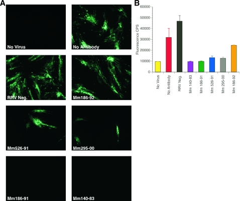
FIG. 7.
Neutralization of RRV-GFP by sera from rhesus monkeys naturally infected with RRV. (A) Fluorescence microscopy of RRV-GFP neutralization. RRV-GFP (MOI of 0.04 PFU/cell) was incubated with either DH20 medium (No Antibody), sera from a RRV-negative rhesus monkey (RRV Neg.) or sera from rhesus monkeys naturally infected with RRV (Mm 186-92, 526-91, 295-00, 186-91, and 140-83). After a 3-h incubation at 37°C, rhesus fibroblasts were inoculated with either DH20 medium (No Virus) or the virus-sera mixture. At 24 h p.i., cultures were rinsed five times with HBSS and refed with DH20. At day 4 p.i., cultures were examined by fluorescence microscopy for GFP expression. (B) Detection of RRV-GFP neutralization by GFP emission intensity. Each bar represents the average GFP counts/s (CPS), with standard deviation, for each rhesus monkey sera tested.

