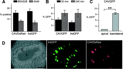
FIG. 1.
Efficacy of CAV-2 vectors in lung tissue. (A) Transduction of human pulmonary cell lines with HAd5 and CAV-2 vectors. The transduction efficiency of CAV-2 and HAd5 vectors was assayed in BEAS2B (epithelial, bronchial) and A549 (epithelial, alveolar) cells. Cells were incubated with CAVDsRed or AdGFP (50 infectious particles/cell) and analyzed by flow cytometry 24 h later. (B) Transduction efficiency of well-differentiated human airway epithelia. Bronchial epithelium was reconstituted in vitro from human primary lung cells after culturing at the medium-airway interface. These cells reconstitute a well-differentiated epithelia, with tight junctions and a basolateral and apical surface. Epithelia were infected with 2.5 × 10³ p.p. of either CAVGFP or AdGFP on the apical surface for 30 or 240 min, and cells were analyzed by FACS 72 h postinfection (n = 8). (C) Polarity of human airway epithelium infection with CAV-2. Epithelia were incubated with 2.5 × 10³ p.p. of CAVGFP on either the apical or basolateral surface for 30 min. Flow cytometry analysis was performed 48 h later (n = 4). **, P < 0.01. (D) Coinstillation in the mouse respiratory tract. CAVDsRed and AdGFP (5 × 10¹⁰ p.p.) were codelivered to 6-week-old C57BL/6 mice by i.n. instillation (n = 4). The lungs were recovered 6 days postinfection. The figure shows an example of a bronchiole in cross-section in which epithelial cells were transduced by CAVDsRed and AdGFP. E, epithelium; L, lumen. Bar, 50 μm.

