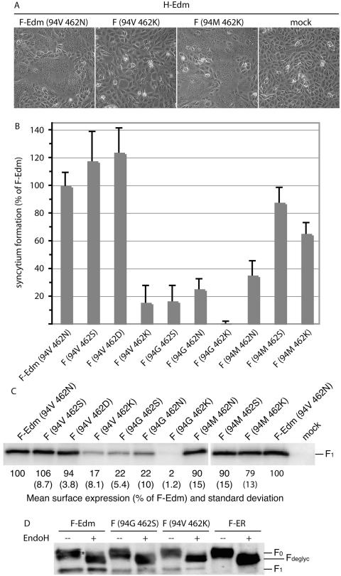
FIG. 4.
The nature of residue 94 in the F cavity determines the effects of changes of residue 462. (A) Syncytium formation after cotransfection of cells with plasmid DNA encoding MV H-Edm and MV F-Edm or F-Edm variants as indicated. Mock-transfected cells (mock) received MV H encoding plasmid only; the plates were photographed 20 h posttransfection at a magnification of ×200 (shown at ×186). (B) Fusogenicity of F-Edm variants cotransfected with H-Edm. Cytotoxicity as an indicator for the ability to induce syncytium formation was quantified as described for Fig. 1B. The values were normalized for unmodified F-Edm and represent the means of three independent experiments. The error bars indicate standard deviations. (C) Surface biotinylation of cells expressing different F-Edm variants to determine F plasma membrane steady-state levels. Biotinylated proteins were precipitated and separated by SDS-PAGE, and F-antigenic material was detected with specific antisera directed against the cytosolic domain of F. The values are based on densitometric quantification using a VersaDoc system and indicate the average percentage of surface material relative to unmodified F-Edm calculated from three to six independent experiments. Standard deviations are shown in parentheses. Mock-transfected cells received vector DNA only. (D) EndoH of F-antigenic material. The F0 fractions of both mutants analyzed were sensitive to EndoH treatment (Fdeglyc material), indicating an ER-type carbohydrate chain conformation. The asterisk marks the small Golgi fraction of EndoH-resistant F-Edm prior to proteolytic maturation. For control, an F variant (F-ER) carrying a KKXX ER retention signal was included. All samples were analyzed by SDS-PAGE and immunoblotting subsequent to EndoH treatment.

