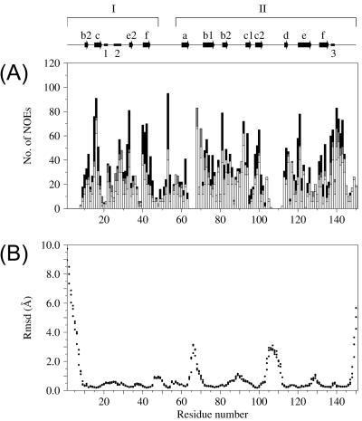
FIG. 2.
Structural parameters for CVB4 2Apro. (A) Distribution (number [No.]) of NOE restraints by residue. From bottom to top, intraresidue (white), sequential (light gray), medium-range (dark gray), and long-range (black) NOEs, respectively. (B) RMS deviations (RMSD) from the average structure for backbone atoms N, Cα, and C′, plotted as separate data points for each residue. The arrows and bars along the top of the figure represent the regions of secondary structure. β-Strands of the N domain (I) are labeled b2, c, e2, and f, whereas those of the C domain (II) are labeled a, b1, b2, c1, c2, d, e, and f. Helical structures are labeled 1, 2, and 3.

