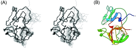
FIG. 3.
3D backbone structure and topology of CVB4 2Apro. (A) Stereoview overlay of the 17 lowest energy structures comprising the structural ensemble. The overlay was performed using the backbone atoms (N, Cα, and C′) of residues 10 to 62, 70 to 102, and 112 to 147. (B) MOLSCRIPT (29) diagram of the representative structure of CVB4 2Apro, shown in the same orientation as for panel A. β-Strands in the N domain (bI2, cI, eI2, and fI) and in the C-terminal domain (aII, bII1, bII2, cII1, cII2, dII, eII, and fII) are labeled following the nomenclature adopted by Petersen et al. (43). The regions exhibiting a helical structure are labeled 1, 2 (α-helix between strands cI and eI2), and 3. The residues comprising the catalytic triad (H21, D38, and C110A) and those involved in zinc ion coordination (C56, C58, C116, and H118) are depicted as wire frames, and the zinc ion is shown as a gray sphere.

