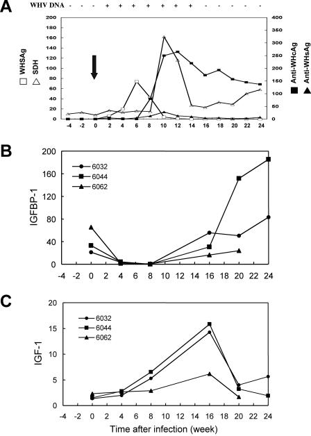
FIG. 8.
Expression of IGFBP-1 and IGF-1 in woodchuck liver infected with WHV. Naïve adult woodchucks were infected with WHV as described previously (38). Blood and liver biopsy samples were collected at various time points before and after the virus inoculation. A representative course of WHV infection with serologic markers and liver function tests is shown (arrow indicates time of infection) (A). Quantitative real-time PCR analysis of the expression for IGFBP-1 (B) and IGF-1 (C) was performed using total RNA isolated from the liver biopsy samples of three separates woodchucks (numbered as 6032, 6044, and 6062) at various time points during acute WHV infection as indicated in the figure. The values of IGFBP-1 and IGF-1 expression were normalized to the level of GAPDH.

