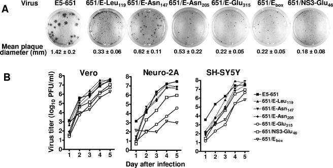
FIG. 2.
Plaque morphology and growth analysis of cDNA-derived E5-651 virus and its recombinant mutants in Vero and neuroblastoma cells. (A) Plaque size of the E5-651 parent and its mutants on Vero cells monolayers. Monolayers of confluent cells were infected with the indicated viruses and incubated at 37°C for 4 days, and resulting plaques were visualized by immunostaining. The value below each well corresponds to the mean plaque diameter in mm ± standard error (n = 10 to 20 plaques per virus). (B) Analysis of growth of parental E5-651 and its recombinant mutant derivatives in Vero cells and in Neuro-2A or SH-SY5Y neuroblastoma cells following incubation at 32°C. Cells were infected with the indicated virus at an MOI of 0.01, and the virus titers in cell culture medium were determined as described in Materials and Methods.

