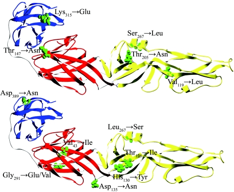
FIG. 3.
Proposed three-dimensional structure of the E protein of LGT E5, based on the structure of the TBEV E protein ectodomain (31). The three structural domains I, II, and III are shown in red, yellow, and blue, respectively. Mutated residues identified in the E protein of the E5-104 mutant (Table 1) are shown in the upper panel. Mutated residues present in virus isolates from brain of mice are shown in the lower panel. The mutant residues are shown in green, and their position numbers and amino acid substitutions are indicated. The structure of the E protein ectodomain is displayed using the Swiss-Pdb Viewer version 3.7 (http://swissmodel.expasy.org/spdbv).

