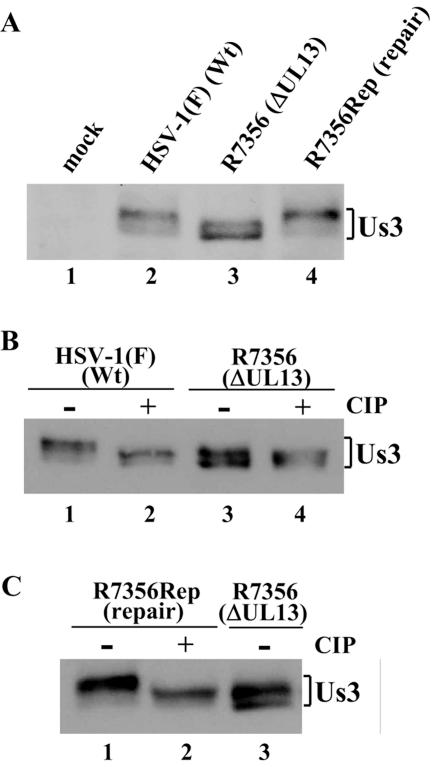
FIG. 1.
(A) Immunoblot of electrophoretically separated lysates from Vero cells mock infected (lane 1) or infected with HSV-1(F) (lane 2), R7356 (lane 3), or R7356Rep (lane 4). Infected cells were harvested at 12 h postinfection and analyzed by immunoblotting with polyclonal antibody to Us3. Wt, wild type. (B) Immunoblots of electrophoretically separated lysates from Vero cells infected with HSV-1(F) (lanes 1 and 2) and R7356 (lanes 3 and 4). The infected cells were harvested at 12 h postinfection, solubilized, mock treated (lanes 1 and 3) or treated with CIP (lanes 2 and 4), and immunoblotted with antibody to Us3. (C) Immunoblots of electrophoretically separated lysates from Vero cells infected with R7356Rep (lanes 1 and 2) and R7356 (lane 3). The infected cells were harvested at 12 h postinfection, solubilized, mock treated (lanes 1 and 3) or treated with CIP (lane 2), and immunoblotted with antibody to Us3.

