FIG. 3.
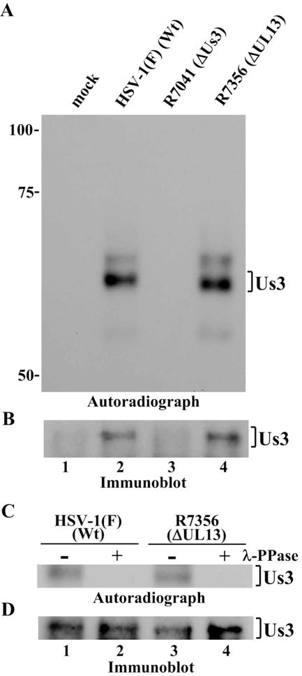
Autoradiographic images of Us3 immunoprecipitates subjected to in vitro kinase assay. (A) Vero cells were mock infected (lane 1) or infected with HSV-1(F) (lane 2), R7041 (lane 3), or R7356 (lane 4); harvested at 12 h postinfection; and immunoprecipitated with antibody to Us3. The immunoprecipitates were incubated in kinase buffer containing [γ-32P]ATP, separated on a denaturing gel, transferred to a nitrocellulose membrane, and analyzed by autoradiography. (B) Immunoblot of the nitrocellulose membrane in panel A using anti-Us3 antibody. (C) Immunoprecipitates prepared as in panel A were either mock treated (lanes 1 and 3) or treated with λ-PPase (lanes 2 and 4), separated on a denaturing gel, transferred to a nitrocellulose membrane, and analzyed by autoradiography. Wt, wild type. (D) Immunoblot of the nitrocellulose membrane in panel C using anti-Us3 antibody.

