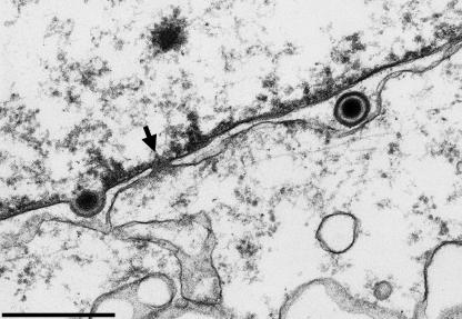
FIG. 1.
Electron micrograph of BHK21 cells 12 h after infection with HSV-1. The arrow denotes an intact nuclear pore next to a capsid undergoing primary envelopment (left) and a primary enveloped virion in the perinuclear space (right). The nuclear side is on the top, and the cytoplasmic side is on the bottom. Bar, 500 nm. (Electron micrograph courtesy of Harald Granzow, Friedrich-Loeffler-Institut, Insel Riems, Germany).

