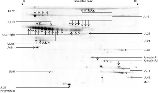
FIG. 2.
Proteins from purified wild-type PrV-Ka virions were separated by two-dimensional electrophoresis, and the resulting protein spots were identified by peptide mass fingerprint analysis. Isoelectric focusing strips were used in the nonlinear gradient range of pH 3 to 10. A number of proteins were found in multiple size and/or charge modifications. Note that isoelectric points of gB isoforms vary from values close to 3 to up to 7. The UL48 protein appears in two horizontal chains of charge variants corresponding to bands 13 and 14 from the one-dimensional analysis (Fig. 1).

