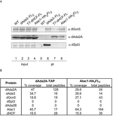
FIG. 3.
Tagging of Atac1, dHCF, and dAda2A confirms their association. (A) S2 cells were transfected with pRmHa3-dAda3-FL2, pRmHa3-Atac1-HA2FL2, and pACXT-T7-dHCF-FLAG plasmids. After a 1-day induction, whole-cell extracts were prepared and incubated with M2-agarose beads. Untransfected S2 cells (WT) were used as a negative control. The immunoprecipitated material was analyzed by Western blotting using antibodies against dGcn5 (α dGcn5, rabbit), dAda2A (α dAda2A, rat), and dSpt3 (α dSpt3, rabbit). Lanes 1 to 4 correspond to 40 μg of whole-cell extract (2% input). Lanes 5 to 8 correspond to the immunoprecipitated material (IP). (B) Protein complexes from a stable line expressing Atac1-HA2FL2 were affinity purified using anti-FLAG-agarose beads. dAda2A-containing complexes were purified from dAda2A-TAP-expressing cells, according to the TAP protocol. These complexes were analyzed by MudPIT. The number of nonredundant spectra for each protein (total peptides) and the amino acid sequence coverage (% coverage) are shown.

