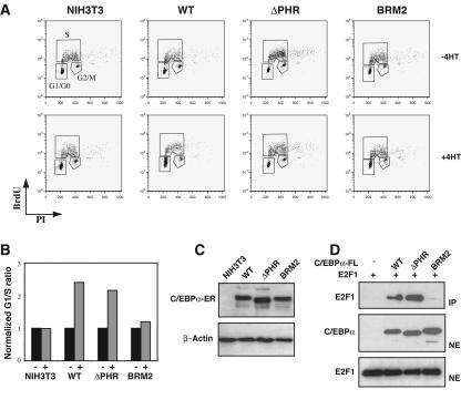
FIG. 2.
In vitro growth repression of C/EBPα-ER fusions. (A) NIH 3T3 clones expressing C/EBPα (WT), C/EBPαΔPHR (ΔPHR), or C/EBPαBRM2 (BRM2). Nuclear localization of C/EBPα-ER or derivatives was accomplished by addition of 1 μM 4-hydroxy tamoxifen (4HT) for 72 h. Following BrdU labeling and propidium iodide staining (see Materials and Methods), cells were subjected to FACS analysis in order to quantify the distribution of cells in the different phases of the cell cycle. The experiments were performed three times. (B) A histogram plot showing the G1/S ratios derived from the experiments shown in panel A. (C) A Western blot of lysates derived from the cells shown in panel A probed with a C/EBPα-specific antibody. A β-actin antibody was used to control for even loading. (D) FLAG-tagged wild-type or mutant C/EBPα was immunoprecipitated from nuclear extracts from cells cotransfected with pcDNA3-C/EBPα-FLAG (WT, ΔPHR, or BRM2) and pCMV-E2F1. Western blot analysis shows E2F1 in the immunoprecipitate (IP) and E2F1 and C/EBPα in the nuclear extracts (NE).

