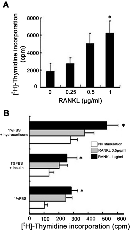
FIG. 1.
RANKL induces the proliferation of primary mammary epithelial cells. (A) Serum-starved primary mammary epithelial cells, isolated from 14.5-day pregnant mice, were stimulated with the indicated doses of RANKL in DMEM containing 10% FBS for 24 h. Cells were incubated with 1 μCi of [3H]thymidine/ml for the last 12 h of culture, and [3H]thymidine incorporation was measured. ✽, significant difference (P < 0.001). (B) Primary mammary epithelial cells isolated from 14.5-day pregnant mice were stimulated with the indicated doses of RANKL in DMEM containing 1% FBS and growth factors for 60 h. Cells were incubated with 1 μCi of [3H]thymidine/ml for the last 24 h of culture, and [3H]thymidine incorporation was measured. The results are shown as mean values ± the standard error of the mean of three separate experiments. ✽, significant difference (P < 0.01).

