FIG. 2.
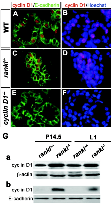
Cyclin D1 expression in rankl−/− mammary glands. (A to F) Immunohistochemistry of cyclin D1 in wild-type (A and B) and rankl−/− (C and D) mammary glands. Tissue sections of mammary glands at 1 day of lactation (L1) from the specified genotypes were stained with anti-cyclin D1 (red)/anti-E-cadherin (green) antibodies (A, C, and E) and Hoechst (B, D, and F). The specificity of antibody staining for cyclin D1 was confirmed by its absence in cyclin D1−/− mammary epithelium (E and F). (G) Western blot analysis of lysates from rankl+/− and rankl−/− mammary tissues at P14.5 and L1. Actin (a) and E-cadherin (b) were used as loading controls. Similar results were obtained from three independent experiments.

