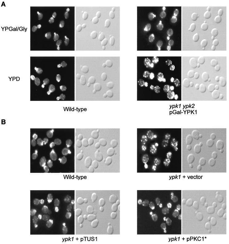
FIG. 5.
ypk cells display an actin organization defect. (A) Wild-type (TB50a) and ypk1 ypk2/pGal-YPK1 (TS57-2B) cells were grown in YPGal/Gly (galactose) medium, shifted to YPD (glucose) medium for 16 h, fixed, stained for actin with TRITC-phalloidin, and observed by fluorescence (actin, left panels) and Nomarski (right panels) microscopy. (B) Overexpression of TUS1 or expression of PKC1* suppresses the actin defect in ypk1 cells. Wild-type (TB50a) cells and ypk1 (TS38-1C) cells transformed with empty vector, pTUS1, or pPKC1* were pregrown in SD medium. Cells were then grown to early-logarithmic phase in YPD medium and processed for actin staining. Fluorescence (actin) and Nomarski microscopy images are shown in the left and right panels, respectively.

