FIG. 6.
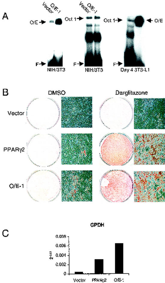
O/E-1 promotes adipocyte differentiation of NIH 3T3 fibroblasts. (A) NIH 3T3 cells were infected with empty retroviruses (vector) or viruses containing O/E-1 or PPARγ2 cDNA. Nuclear extracts were prepared, and the presence of O/E protein was analyzed by EMSA with the mb-1 O/E-1 probe. The Oct probe was included as a control for protein content, and nuclear extracts from 3T3-L1 cells, differentiated for 4 days by the addition of MDI, were included for a comparison of O/E levels. F indicates free probe. (B) Infected NIH 3T3 cells were treated with MDI and either darglitazone or the vehicle (DMSO) 2 days after confluence. The cultures were stained with Oil Red O after 10 days of differentiation. Petri dishes and micrographs from one representative experiment out of four are shown. (C) GPDH levels of infected cells after differentiation. Infected cells were induced to differentiate by the addition of MDI and darglitazone, and RNA samples were harvested after 4 days of differentiation. DNase-treated samples were used for cDNA synthesis and subsequent quantitative real-time-PCR analysis. The results of one representative experiment out of two are shown.

