FIG. 6.
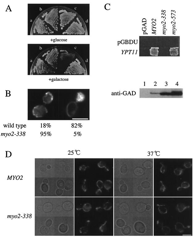
Phenotypes of myo2-338 cells. (A) Effect of overexpression of YPT11 on myo2-338 cells. myo2-338 cells (strain yTO014 [a and b]) and wild-type cells (strain YPH499 [c and d]) with YIpUGAL7-YPT11 (b and c), YPT11 under the control of the GAL7 promoter, or without YIpUGAL7-YPT11 (a and d) were streaked on YPD (+glucose) and YPG (+galactose) plates and incubated at 30°C for 3 days. (B) Mitochondrial distribution in myo2-338 cells overexpressing YPT11. Wild-type cells (strain YPH499) and myo2-338 cells (strain yTO014) with YIpGAL7-YPT11 were cultured until early log phase in SC raffinose, shifted into SCGal medium for induction, incubated for 4 h at 25°C, stained with DASPMI, and observed to count the number of cells with a normal distribution of mitochondria (left) or with mitochondria accumulated in the bud (right). More than 200 of cells with a middle-sized bud were observed. (C) Two-hybrid interaction of mutant Myo2p with Ypt11p. (Top) Reporter cells (strain PJ69-4A) were transformed with pGBDU-C1 (pGBDU) for a control or pK027, a pGBDU-C1-based plasmid for Ypt11p fused with the UASGAL-binding domain (YPT11), and pGAD-C1-based constructs of MYO2 (pK016 for MYO2, pK017 for myo2-338, pK018 for myo2-573) or pGAD-C1 for a control (pGAD). The cells were streaked on SC medium lacking uracil, leucine, histidine, and adenine, where two-hybrid interactions were detected as cell growth. (Bottom) Relative amounts of the fusion proteins of Myo2p with the trans-activator domain (GAD) in the cells above were analyzed by Western blotting using anti-GAD antibodies (lane 1, control; lane 2, cells with pK016, lane 3, cells with pK017, lane 4, cells with pK018). myo2-338 from the pGAD-based construct was expressed more strongly than MYO2 from the pGAD-based construct; however, two-hybrid interaction was not detected. (D) Morphology of mitochondria in myo2-338 cells. Wild-type cells (strain YPH499, MYO2) and myo2-338 cells (strain yTO014, myo2-338), producing mitochondrion-targeting GFP, were cultured until early log phase at 25°C (25°C), shifted to 37°C, and incubated for 2 h (37°C). Phase-contrast images (left) and GFP signals (right) of each culture are shown. Bars, 5 μm.

