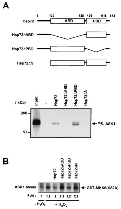
FIG. 6.
Interaction of ASK1 with the NH2-terminal region and the ABD of Hsp72. (A) In vitro-translated 35S-labeled full-length ASK1 was applied to hexahistidine-tagged Hsp72 variants that had been immobilized to Ni2+-NTA-agarose beads. Bead-bound proteins were subsequently eluted and analyzed by SDS-PAGE and autoradiography as for Fig. 5A. The input 35S-labeled ASK1 (20%) is also shown. A schematic diagram of Hsp72 and its mutants is shown above the gel. (B) NIH 3T3 cells were transiently transfected for 48 h with pcDNA3-Flag-ASK1 and were then incubated for 20 min at 37°C in the absence or presence of 2 mM H2O2. Cell lysates were subjected to immunoprecipitation with an anti-Flag antibody. The resulting precipitates were incubated for 1 h at room temperature with 2 μg of purified wild-type Hsp72, Hsp72ΔABD, Hsp72ΔPBD, or Hsp72ΔN in 50 μl of HEPES buffer (pH 7.4), washed twice with the HEPES buffer, and then assayed for ASK1 activity with GST-MKK6(K82A) as the substrate.

