FIG. 4.
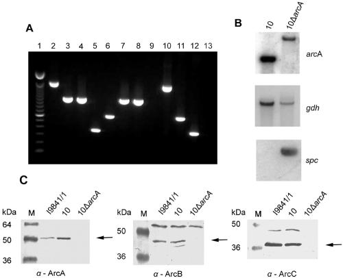
Construction of an arcA knockout mutant. (A) Control of strain 10ΔarcA in comparison with wild-type strain 10 by PCR. An ethidium bromide-stained agarose gel with the PCR DNA fragments is shown. Amplification was performed with chromosomal DNAs of strain 10ΔarcA (lanes 2 to 5) and wild-type strain 10 (lanes 6 to 9). ΔarcA-containing plasmid pICADspc (lane 10), wild-type arcA-containing plasmid pBAD (lane 11), and spectinomycin donor plasmid pICspc (lane 12) were used as positive controls, and double-distilled H2O (lane 13) was used as a negative control. Lane 1, molecular weight marker; lanes 2, 6, and 10, amplification reaction to verify insertion of the spc resistance gene cassette (1,609-bp fragment, successful insertion; 614-bp fragment, no insertion); lanes 3 and 7, arcB-specific DNA fragment (1,039 bp); lanes 4 and 8, arcC-specific DNA fragment (1,027 bp); lanes 5, 9, and 12, spc cassette-specific DNA fragment (361 bp). (B) Southern analysis of wild-type strain 10 and mutant strain 10ΔarcA with probes specific for the arcA, gdh (41), and spc genes. (C) Immunoblot analyses with whole-cell lysates of S. suis strains I9841/1, 10, and 10ΔarcA (lane M, molecular size marker). Specific antisera against the ArcA, ArcB, and ArcC (α-ArcA to -C) proteins were used at a 1:200 dilution. The specific protein bands are indicated by arrows.

