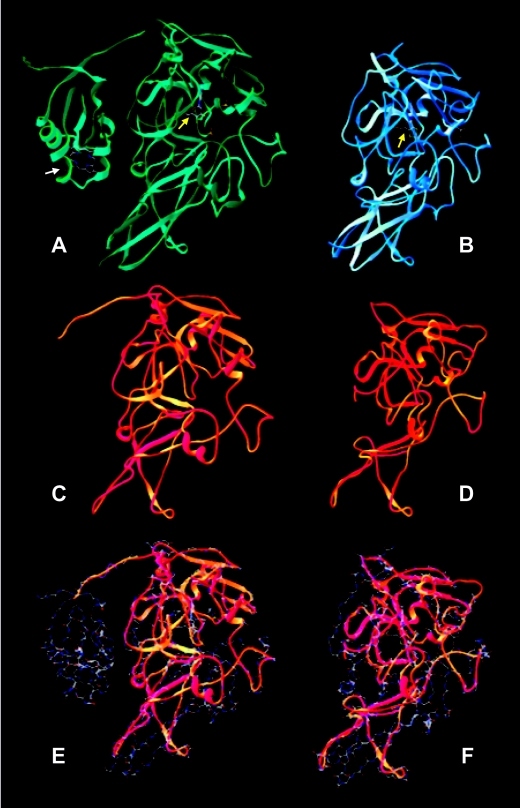
FIG. 2.
Structural prediction for DraSO based on A. thaliana and chicken liver SO models. Superimposition models with both the available chicken SO and plant SO (At-SO) structures were obtained with the Deep View Swiss-PdbViewer, version 3.7. (A and B) Three-dimensional structures of bird and plant SOs. The view shows the heme binding domain in chicken SO (indicated by a white arrow) that is absent in At-SO and the binding to molybdopterin coenzyme (indicated by yellow arrows) in both structures. (C and D) Predicted structure of DraSO using chicken SO and At-SO as superimposition models, respectively. (D and F) Superposition of DraSO (ribbons) on chicken SO and At-SO (wire frame).

