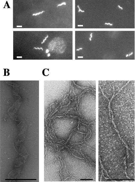
FIG. 3.
Scc forms polymeric structures in vitro. (A) Fluorescence microscopy imaging of polymers of L. biflexa Scc fused to GFP. Typical filaments are 2 to 3 μm long. Scale bar, 1 μm. (B and C) Electron microscopy of in vitro self-assembly of Scc-6His alone. Helix-like structures (B) and filaments with a cross-sectional diameter of 6 to 10 nm (C) were observed. Similar pictures were observed for Scc-GFP. Purified recombinant Scc-GFP or Scc-6His protein (1 μg/μl) was incubated 1 h at 30°C in 50 mM Tris, pH 7.5, and 150 mM NaCl. Scale bar, 500 nm (B) or 100 nm (C).

