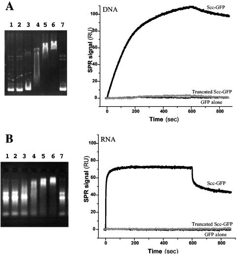
FIG. 6.
Nucleic acid binding assays of Scc. (Left) Electrophoretic mobility shift assays of L. biflexa Scc. Agarose gel electrophoresis staining with ethidium bromide for visualizing DNA (A) and RNA (B). Lanes 1 and 7, 1 μg plasmid DNA (A) or 1 μg total RNA from L. biflexa (B). Lanes 2 to 6, 1 μg of plasmid DNA (A) or total RNA (B) with 2, 5, 10, 20, and 40 μg of Scc-6His protein. All reactions were carried out in a total volume of 40 μl. (Right) Real-time surface plasmon resonance (SPR) profiles showing the interaction between immobilized L. biflexa Scc-GFP-6His and 250-μg/ml plasmid DNA (A) or 150-μg/ml total RNA (B) in solution. No binding was observed on GFP or truncated Scc (95-212)-GFP-6His.

