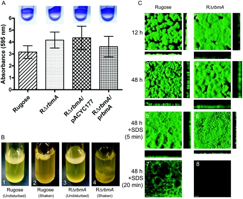
FIG. 4.
RbmA is required for biofilm structure. (A) Quantitative comparison of biofilm formation by the wild-type rugose variant, RΔrbmA, RΔrbmA/pACYC177, and RΔrbmA/prbmA after an 8-h incubation in LB medium at 30°C under static conditions. Error bars represent standard deviations. Crystal violet-stained biofilms formed in the wells of polyvinyl chloride microtiter plates, which were used in the quantitative biofilm assays, are shown in the inserts. (B) Pellicle formation by the wild-type rugose variant (B1 and B2) and RΔrbmA (B3 and B4) after 2 days of incubation in LB at 30°C. B2 and B4 show pellicles disturbed by shaking. (C) Confocal scanning laser microscopic images of horizontal (xy) and vertical (xz) projections of biofilm structures formed by the rugose variant and RΔrbmA in a flow cell system. Biofilm structures that developed after 12 and 24 h of incubation are shown in panels C1 to C4. Biofilm structure integrity studies using SDS are shown in C5 to C8, and the duration of SDS treatment is indicated on the left. In C1 and C2, bars represent 30 μm, whereas in C3 to C8, they represent 100 μm.

