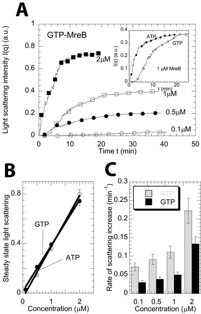
FIG. 2.
Assembly of ATP-MreB and GTP-MreB. (A) Time-dependent light-scattering intensity for GTP-MreB assembly. Light scattering was measured at an angle of 90° from the direction of the incident light. Symbols correspond to 0.1 μM (○), 0.5 μM (•), 1 μM (□), and 2 μM (▪) GTP-MreB, respectively. The inset shows the intensity versus time for MreB in the presence of ATP (•) and GTP (○). (B) Steady-state light-scattering intensity as a function of total concentration of protein in solution for ATP-MreB and GTP-MreB. (C) Rates of increase of light scattering of MreB in the presence of ATP and GTP. The rate was calculated as the inverse of time it takes to reach 90% of its steady-state value.

