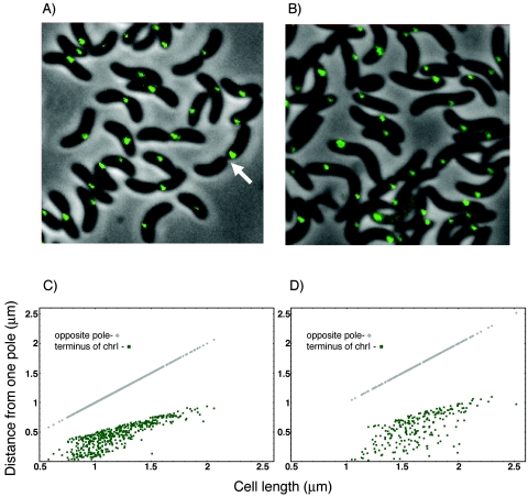
FIG. 3.
Localization of the terminus region of chrI marked with λOL1 operators. Shown are representative fields of L broth (A)- and M63-plus-glucose (B)-grown cells (CVC244) showing a single fluorescent focus even when invagination is well advanced (arrow). Distribution of focus positions in cells under the two growth conditions is shown in panels C and D, respectively. The focal distance was measured from the cell pole, which was closer to the focus.

