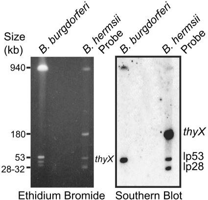
FIG. 2.
Southern blot analysis of replicons of B. hermsii and B. burgdorferi separated by pulsed-field gel electrophoresis and sequentially hybridized with radiolabeled probes for thyX, the lp53 linear plasmid, and the lp28 linear plasmid of B. hermsii. An ethidium bromide-stained gel is shown on the left, and an autoradiograph is shown on the right. Sizes (in kb) of linear plasmids of B. hermsii and of size standards are indicated.

