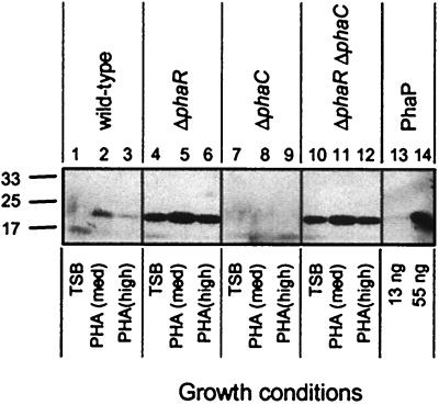
FIG. 1.
Immunoblots of PhaP in R. eutropha strains. Proteins were separated by sodium dodecyl sulfate-polyacrylamide gel electrophoresis (SDS-PAGE) and were subjected to immunoblot analysis for detection of PhaP. Cells from R. eutropha cultures were harvested after cultivation for 48 h in TSB, PHA(med), or PHA(high). Bacterial samples correspond to cells from 10 μl of a culture diluted to an OD600 of 0.2. Purified PhaP was included as a control. Molecular mass standards are indicated in kilodaltons. Culture OD600 measurements were as follows: lane 1, 8.9; lane 2, 8.3; lane 3, 9.6; lane 4, 8.6; lane 5, 7.2; lane 6, 5.7; lane 7, 9.1; lane 8, 4.3; lane 9, 1.3; lane 10, 8.9; lane11, 3.8; lane 12, 1.8.

