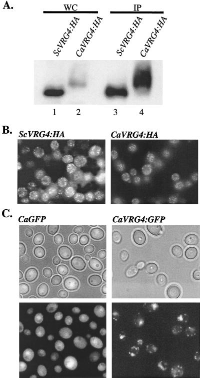
FIG. 3.
Expression and intracellular localization of CaVrg4p. (A) Western blot analysis of CaVrg4-HA and ScVrg4-HA proteins. Whole-cell protein extracts were prepared from S. cerevisiae strains that were grown in YPAD to repress the endogenous GAL1p-VRG4 gene (XGY14) (25) and harbored plasmids expressing either ScVRG4-HA (pTiVRG4-HA) (25) or CaVRG4-HA (pTiM1-HA). Equivalent amounts of proteins were directly applied to SDS-polyacrylamide gels (WC) (lanes 1 and 2) or were first immunoprecipitated with anti-HA mouse antibodies (IP) (lanes 3 and 4), and then the proteins were Western blotted with anti-HA rabbit antibodies and detected by chemiluminescence as described in Materials and Methods. (B) Indirect immunofluorescence of S. cerevisiae strains expressing ScVRG4-HA or CaVRG4-HA. S. cerevisiae strain XGY14 expressing ScVRG4-HA or CaVRG4-HA (as described above) was grown in YPAD to the log phase. Cells were fixed and treated with anti-HA antibodies, followed by fluorescein isothiocyanate-conjugated anti-mouse immunoglobulin G. (C) CaVRG4-GFP (pCaV:GFP) or GFP alone (pAW6) was expressed in C. albicans strain ANC2, and live cells were examined by fluorescence microscopy.

